Tuberculosis is one of the most common treatable infectious disease in India. Most common presentation being pulmonary tuberculosis. Primary tuberculosis of maxillary sinus leading to chronic otitis media is an extremely rare entity, diagnosis of which is often delayed and resulting in delay in start of actual treatment. The following report highlight a case of 63 years male patient presented with bilateral ear discharge, not responding to the routine antibiotic therapy. CT scan imaging denoted left pansinusitis with osteomyelitic changes in left maxillary sinus with otitis media. The diagnosis was confirmed by histopathological biopsy report. The patient was treated with Anti-tubercular regimen for 9 months and following which bilateral tympano-mastoidectomy was done.
On examination, the patient had bilateral subtotal perforation and bilateral persistent ear discharge. Discharge was slightly mucoid, non-foulsmelling and continuous. An ear swab was taken for culture sensitivity, which did not denote any organism growth.
Routine blood examination showed reduced haemoglobin level, red blood cell count and platelet count whereas erythrocyte sedimentation rate and white blood cell count were increased. Chest X-ray PA view shows normal study.
CT scan of temporal bone showed bilateral opacification with soft tissue in the mastoid air cells and in the aditus-ad-antrum region.
A pure tone audiogram showed bilateral moderate conductive hearing loss.
CT scan paranasal sinus showed left-sided pansinusitis with osteomyelitic changes in the left maxillary sinus.
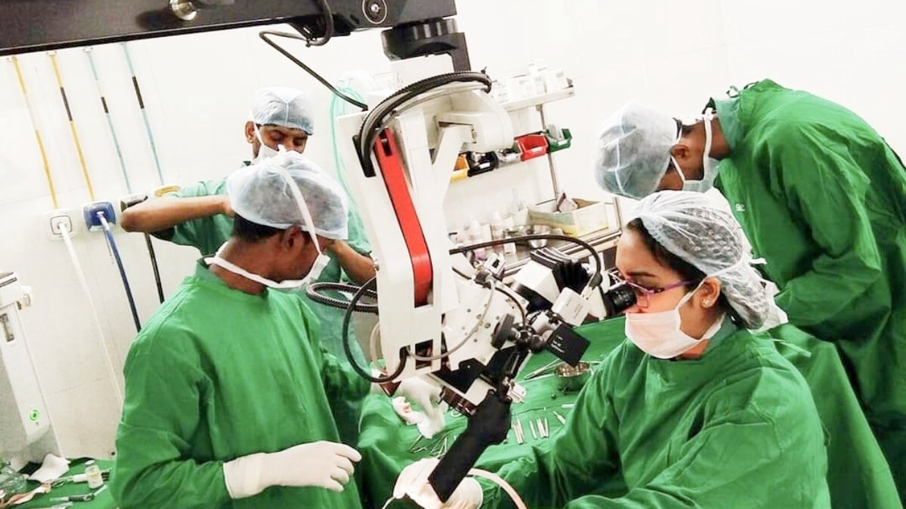
The patient was operated on for functional endoscopic sinus surgery. Uncinectomywith type III maxillary antrostomy was done, necrotic tissue with purulent secretion in the left maxillary sinus was removed. Osteomyelitic changes were seen in the anteroinferior bony wall of the maxillary antrum.
On completion of surgery on the left side, nasal washing of all the sinuses was done.
Incidentally, the patient had a maxillary sinus polyp in the right maxillary sinus which was removed.
Tissue from left maxillary sinus sent for histopathological evaluation which revealed inflammatory granulation tissue and necrotizing epithelioid granulomas with a large area of the caseous type of necrosis with entrapped bone spicules suggestive of tuberculous infection. Pus from left maxillary sinus taken and send for Ziehl-Neelson staining which showed the presence of Mycobacterium tuberculosis.
The patient started on standard anti-tubercular medications comprising of 4 drugs isoniazid 5 mg/kg/day, rifampin 10 mg/kg/day, ethambutol 15 mg/kg/day, pyrazinamide 25 mg/kg/day starting with an intensive phase of 2 months, followed by a continuation phase of 7 months.
Image 1
Image 2
Within 6-8 weeks of anti-tubercular treatment bilateral ear discharge subsided. The patient completed the standard anti-tubercular treatment for 9 months. The patient was operated on for right tympano-mastoidectomy 5 months after commencement of the anti-tubercular drugs. During ear surgery, the middle ear mucosa was slightly pale. Aditus patency was achieved, ossicular assembly checked and tympanic membrane grafting was done by 270-degree cuff technique using temporalis fascia. Biopsy of the mucosa from the mastoid antrum did not reveal any active infection.
A similar operation is done on the left ear 8 months after commencement of the anti-tubercular drugs. A post-operative pure tone audiogram did 6 months following surgery showed an air-bone gap between 10 to 20 dB.
Image 3
Regular follow up and diagnostic nasal endoscopy done every 3 months for 1 year then every 6 months for 2 years did not show any evidence of disease. There was good mucosalization of the paranasal sinus cavity with bilateral tympanic membrane graft uptake with hearing improvement.
DISCUSSION
Incidence of tuberculosis reduced significantly in the twentieth century, but drug-resistant anti-tubercular strains and the immunodeficient host has lead to a re-emergence of tuberculosis. Extra-pulmonary tuberculosis amounts to 20 per cent of total tuberculosis cases. Giovanni Morganifirst reported nasal tuberculosis in the year 1761. He classified nasal tuberculosis as spontaneous nasal tuberculosis (occurs secondarily after pulmonary tuberculosis) and primary nasal tuberculosis (occurs without primary pulmonary tuberculosis). Infection of the middle ear occurs by the passage of infected material from the nasal cavity and nasopharynx via eustachian tube while coughing or sneezing or by hematogenous spread, Nasal tuberculosis may present with nasal obstruction, nasal discharge, epistaxis and crust formation of the middle ear. Tuberculosis of the middle ear leads to painless persistent otorrhoea, often multiple tympanic membrane perforations, bone erosion, granulation of the middle ear and mastoid and if no treatment is given then there is rapid extensive destruction of the middle ear and its.
surroundings. Pure tone audiogram shows a conductive hearing loss, nevertheless, sensorineural hearing loss occurs when labyrinth is involved. Definitive diagnosis is made by taking nasal discharge and tissue for acid-fast bacilli or the presence of a typical tuberculous nodule. Nowadays polymerase chain reaction (PCR)is used to make diagnosis of tuberculosis. But in some cases, PCR may give a negative value, while tissue biopsy is positive. Hence PCR and tissue biopsy should complement each other to make a final diagnosis.
The primary treatment of tuberculosis is anti-tubercular medication, till the patient gets free of symptoms of tuberculosis, followed by secondary treatment if required.
In our case, an elderly male patient presented with bilateral ear discharge as the presenting symptom without any nasal symptoms. Nasal involvement was suspected as ear discharge was not responding to the routine course of medical therapy. On investigation of CT scan, paranasal sinus and culture of pus concluded that patient had isolated tuberculosis of the left maxillary sinus with pansinusitis.
Though the organisms could not be isolated on the ear swab culture, the possibility of the middle ear effusion having a tubercular infection is likely as the patient immediately responded to the anti-tubercular drug regimen. Surgery for the ear was done 5 months after commencement of the anti-tubercular drug treatment and clinical improvement.
This patient had a post-operative hearing improvement and no recurrence was seen in 2 year follow up.
Juvekar M et al. Int J Otorhinolaryngol Head Neck Surg. 2020 Aug;6(8):1576-1578International Journal of Otorhinolaryngology and Head and Neck Surgery.
Source: https://www.ijorl.com/index.php/ijorl/article/view/2379
- Department of Otorhinolaryngology, Grant Medical College and J.J Group of Hospitals and Bombay Hospital and
Research Centre, Mumbai, Maharashtra, India. - Department of Otorhinolaryngology, Guwahati Neurological Research Centre, Kolkata, West Bengal, India.